The evolution of modern medicine and industrial inspection owes much of its progress to the development and refinement of X Ray Machines. These powerful diagnostic tools have transformed from experimental curiosities in the late 19th century into essential equipment that defines the standards of modern healthcare and security. By capturing internal structures without the need for invasive procedures, this technology provides clarity where the human eye fails, acting as the foundation for accurate diagnoses and precise quality control across global industries.
The Fundamental Principles of X-Ray Technology
At their core, X Ray Machines operate by emitting electromagnetic radiation at high frequencies. When these rays pass through an object—be it a human limb or a manufactured component—different densities absorb the radiation at varying rates. Bone, for instance, is dense and absorbs a significant amount of radiation, appearing white on the resulting image. In contrast, soft tissues or air gaps allow more rays to pass through, resulting in darker areas. This fundamental interaction between radiation and matter allows for the detailed visualization required by radiologists and engineers alike.
Modern equipment has significantly improved the safety profile of these diagnostic tools. Through the implementation of digital imaging sensors and refined collimation techniques, X Ray Machines can now capture high-resolution images while utilizing lower doses of radiation compared to older film-based counterparts. This advancement has made routine checkups safer and more efficient for patients and operators.
Varieties of X-Ray Systems in Use Today
Depending on the industry, the configuration of the machinery varies significantly. Medical facilities require different specifications than airports or industrial warehouses. Understanding these differences is crucial for selecting the right technology for specific applications:
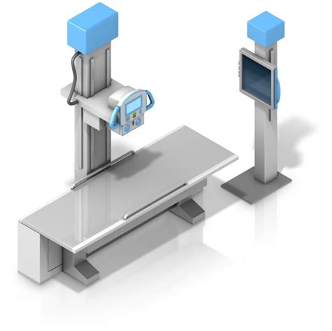
- Digital Radiography (DR): The gold standard in modern hospitals, offering near-instant image acquisition on digital screens.
- Computed Tomography (CT) Scanners: Utilize rotating gantries to create 3D cross-sectional views of the body, providing much more detail than 2D scans.
- Fluoroscopy Systems: Used for real-time visualization, allowing surgeons to watch moving structures like the heart or digestive system during procedures.
- Security Screening Units: Employed in transportation hubs to analyze luggage contents for prohibited items through advanced material discrimination software.
- Industrial Inspection Units: Used in manufacturing to detect cracks, voids, or assembly errors in metallic parts, electronics, and aerospace components.
⚠️ Note: Always ensure that all X-ray equipment is shielded and calibrated by certified professionals to minimize occupational radiation exposure and maintain image quality.
Comparative Analysis of Diagnostic Systems
The following table illustrates the common differences between various imaging platforms used in clinical environments today.
| System Type | Best Used For | Image Format |
|---|---|---|
| Standard Radiography | Bone fractures and chest conditions | 2D Static |
| Fluoroscopy | Real-time internal motion | Video/Dynamic |
| CT Scanners | Complex organ/tissue mapping | 3D Volumetric |
| Dental X-Ray | Oral and jaw assessment | 2D/Panoramic |
Safety Protocols and Maintenance
Operating X Ray Machines is a task that demands rigorous adherence to safety standards. Because ionizing radiation carries inherent risks, the focus is always on the ALARA principle—keeping exposure As Low As Reasonably Achievable. This involves not only the design of the machine itself but also the environment in which it operates. Lead-lined walls, protective aprons, and periodic shielding inspections are mandatory requirements for any facility utilizing this technology.
Maintenance is equally critical. Calibration ensures that the beam quality remains consistent over time, preventing degradation in image sharpness. When a machine begins to drift from its factory settings, diagnostic errors can occur, which could lead to missed medical conditions or structural failures in industrial parts. Therefore, a proactive maintenance schedule is the most effective way to protect the longevity of the equipment and the safety of the user.
💡 Note: Regularly inspect all personal protective equipment (PPE), such as lead aprons, for cracks or damage that might allow radiation leakage.
The Future of Diagnostic Imaging
The next generation of X Ray Machines is moving rapidly toward Artificial Intelligence integration. Advanced software algorithms are currently being developed to assist radiologists by automatically highlighting potential abnormalities in medical scans. This does not replace the expert eye of the physician but acts as a secondary layer of scrutiny that increases accuracy and reduces burnout. Furthermore, portable and mobile X-ray units are becoming more powerful, allowing emergency medical teams to perform high-quality diagnostic imaging directly at the point of care, significantly improving outcomes in trauma cases.
As we look forward, the trend is toward miniaturization and enhanced connectivity. Wireless sensors allow images to be sent instantly to cloud-based diagnostic platforms, facilitating remote consultations between experts regardless of geographical boundaries. By lowering the barrier to access and increasing the precision of the hardware, the impact of these systems on global health and safety continues to grow, ensuring that we can solve complex structural problems with speed and reliability.
In summary, the role of these imaging systems is indispensable in both the medical and industrial landscapes. By leveraging the physical properties of radiation to observe the unseen, we enable earlier diagnoses, safer surgical interventions, and higher standards of material manufacturing. Whether it is a digital radiography unit in a bustling metropolitan hospital or a rugged security scanner at a global logistics hub, the technology behind these machines serves as a quiet but powerful backbone to modern society. Continuous innovation in hardware and software ensures that these tools remain efficient, safe, and increasingly accessible to those who need them most.
Related Terms:
- radiology machine
- x ray machine images
- x ray equipment
- x ray machine diagram
- x ray machine uses
- digital x ray machine