When it comes to advanced medical diagnostics, the MRI imaging machine stands out as one of the most sophisticated and essential tools in modern healthcare. By utilizing powerful magnetic fields, radio waves, and intricate computer processing, this technology generates highly detailed images of the organs and tissues within the body. Unlike X-rays or CT scans, which rely on ionizing radiation, magnetic resonance imaging provides a safe, non-invasive method for physicians to diagnose a wide array of conditions, ranging from ligament tears and brain tumors to complex cardiovascular issues.

How an MRI Imaging Machine Works
At its core, the MRI imaging machine operates on the principles of nuclear magnetic resonance. Inside the machine is a giant, powerful magnet that creates a steady, strong magnetic field. When a patient enters the scanner, the hydrogen atoms in their body—which are abundant in water and fat—align with this magnetic field.
The machine then emits radio frequency waves that knock these hydrogen atoms out of their aligned position. When the radio waves are turned off, the atoms return to their original alignment, releasing energy in the process. Sensitive receivers in the scanner detect these signals, and a computer converts them into the high-resolution cross-sectional images that radiologists interpret. This entire process is incredibly precise, allowing for the differentiation between healthy and diseased tissue with remarkable accuracy.
Key Applications of MRI Technology
Because of its exceptional soft-tissue contrast, the MRI imaging machine is the gold standard for imaging many parts of the body. Its versatility makes it indispensable across various medical specialties.
- Neurology: It is highly effective for imaging the brain and spinal cord, detecting conditions such as strokes, multiple sclerosis, tumors, and aneurysms.
- Orthopedics: Surgeons rely on MRI to examine joints, muscles, ligaments, and tendons, making it crucial for identifying sports injuries like torn ACLs or rotator cuff damage.
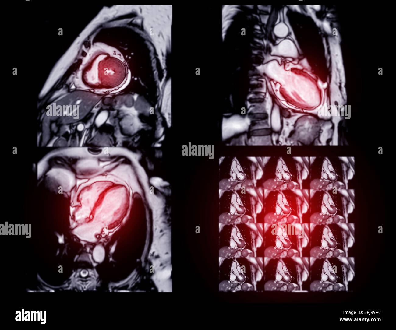
- Cardiology: Cardiac MRI provides detailed assessments of the heart structure, function, and blood flow, helping to diagnose congenital heart defects and damage from heart attacks.
- Oncology: It is instrumental in detecting, staging, and monitoring tumors throughout the body, as well as evaluating the success of cancer treatments.
- Abdominal and Pelvic Imaging: Doctors use it to examine the liver, kidneys, uterus, ovaries, and prostate for abnormalities.
Comparing Diagnostic Imaging Modalities
Understanding when to use an MRI imaging machine versus other modalities like CT scans or X-rays is vital for effective patient care. The following table highlights the primary differences to help clinicians and patients understand these tools better.
| Feature | MRI Imaging Machine | CT Scan | X-Ray |
|---|---|---|---|
| Technology | Magnetic Fields/Radio Waves | X-rays (Ionizing Radiation) | X-rays (Ionizing Radiation) |
| Primary Use | Soft Tissue/Neurological | Bone/Organ/Trauma | Fractures/Chest Imaging |
| Scan Time | Longer (30-60 mins) | Very Fast (Minutes) | Fastest (Seconds) |
| Resolution | Extremely High (Soft Tissue) | High (Bone/Organ) | Lower (General) |
⚠️ Note: Patients with certain metal implants, such as pacemakers, cochlear implants, or specific aneurysm clips, may not be able to undergo an MRI due to the powerful magnetic field. Always inform the radiology staff of any surgical history or metallic objects in your body prior to the scan.
Patient Preparation and What to Expect
Preparing for an appointment with an MRI imaging machine is generally straightforward, but it requires careful attention to safety protocols. Since the magnet is always active, safety is the top priority for both the patient and the staff.
Before the procedure, you will be asked to complete a screening questionnaire to ensure you do not have any contraindicated metal objects. You must remove all jewelry, watches, hearing aids, and hairpins. Some procedures may require a contrast agent, known as gadolinium, which is injected intravenously to improve the visibility of specific tissues or blood vessels.
During the scan, it is essential to remain as still as possible. The MRI imaging machine makes loud tapping, banging, or whirring noises as the internal gradient coils vibrate; this is a normal part of the imaging process. Staff will typically provide earplugs or noise-canceling headphones to minimize discomfort, and you will have an intercom system to communicate with the technologist throughout the duration of the scan.
💡 Note: If you suffer from claustrophobia, inform your physician in advance. They may be able to prescribe a mild sedative to help you remain calm and still, or they may suggest a "wide-bore" or "open" MRI machine if one is available in your area.
Advancements in Magnetic Resonance Imaging
The field of radiology is constantly evolving, and recent advancements have significantly enhanced the capabilities of the MRI imaging machine. Modern scanners now offer higher magnetic field strengths (such as 3 Tesla and above), which produce clearer images in less time. Additionally, functional MRI (fMRI) allows doctors to map brain activity by tracking changes in blood flow, providing insights into cognitive functions and mapping areas responsible for critical movements or speech.
Another major leap is the development of artificial intelligence (AI) integration within the software of the MRI imaging machine. AI algorithms can help reduce background noise, speed up image acquisition, and assist radiologists in identifying subtle abnormalities that might otherwise be overlooked. These innovations continue to push the boundaries of what is possible in diagnostic medicine, leading to faster results and more accurate treatment plans for patients worldwide.
The role of the MRI imaging machine in modern medicine cannot be overstated. By providing a clear, non-invasive window into the internal structures of the human body, this technology serves as a cornerstone for accurate diagnosis and effective long-term treatment planning. As scanning speeds improve, patient comfort becomes a greater focus, and AI-driven software continues to enhance image quality, the future of magnetic resonance imaging looks increasingly bright. Ultimately, these advancements ensure that physicians have the most reliable data possible to make life-changing decisions, emphasizing the vital importance of this medical technology in maintaining global health standards.
Related Terms:
- list of mri devices
- 4 types of mri machine
- mri scan machine types
- which mri machine is best
- 2 types of mri
- 3 types of mri machines