The coxal hip bone, frequently referred to in anatomical terms as the os coxae or the innominate bone, serves as the critical structural foundation for the human pelvis. It is a large, irregularly shaped bone that connects the axial skeleton to the lower limbs, facilitating essential functions such as weight-bearing, locomotion, and the protection of internal pelvic organs. Understanding the anatomy of this complex bone is fundamental for medical professionals, students, and anyone interested in human biomechanics, as it bridges the gap between the torso and the legs.
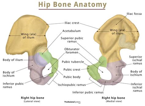
Anatomy of the Coxal Hip Bone
The coxal hip bone is not a single solid structure in adults but is rather formed by the fusion of three distinct bones that coalesce during late adolescence. These three components—the ilium, the ischium, and the pubis—meet at a deep, cup-shaped socket called the acetabulum, which articulates with the head of the femur to form the hip joint.
Each component brings unique structural contributions to the pelvic girdle:
- Ilium: The largest and most superior portion, forming the prominent "hip bone" that can be felt along the waistline. It provides a broad surface for the attachment of major muscles of the trunk and lower extremities.
- Ischium: The posteroinferior part of the bone. It is the portion of the pelvis we sit upon; the ischial tuberosity is particularly known as the "sit bone."
- Pubis: The anteroinferior portion that meets its counterpart from the opposite side at the pubic symphysis, a cartilaginous joint that provides structural stability.
Structural Functions and Biomechanics
The primary role of the coxal hip bone is to act as a weight-bearing structure. When standing, the weight of the upper body is transferred from the vertebral column to the sacrum, and then through the sacroiliac joint to the ilium, ultimately distributing the force to the lower limbs through the acetabulum. This mechanism is crucial for bipedal movement.
Beyond weight distribution, the bone acts as a protective shield for the pelvic viscera, including the urinary bladder, parts of the large intestine, and, in females, the reproductive organs. Furthermore, the extensive surface area of the iliac crest and other bony landmarks serves as an attachment point for various muscles, including the gluteus muscles, which are vital for hip stabilization, walking, and running.
| Component | Location | Primary Function |
|---|---|---|
| Ilium | Superior (Top) | Weight transfer and muscle attachment |
| Ischium | Posteroinferior (Back/Bottom) | Supports body weight while sitting |
| Pubis | Anteroinferior (Front/Bottom) | Protects pelvic organs and forms joints |
💡 Note: While the coxal bone is fused in adults, it originates from cartilage in infancy, which is why pediatric pelvic assessments focus on different growth markers compared to adult clinical examinations.
Clinical Significance of the Hip Region
Because the coxal hip bone is a primary weight-bearing junction, it is susceptible to various clinical issues. Fractures of the pelvis, often resulting from high-impact trauma like motor vehicle accidents, can be life-threatening due to the proximity of major blood vessels and organs. Additionally, degenerative conditions such as osteoarthritis of the hip joint, where the cartilage in the acetabulum wears down, can significantly limit mobility and cause chronic pain.
Physiotherapists and orthopedic specialists often analyze the alignment of the coxal hip bone to address issues related to gait (how a person walks) or chronic lower back pain. Misalignment or pelvic tilt can cause uneven distribution of force throughout the body, leading to secondary issues in the knees and ankles. Maintaining pelvic health through strength training and flexibility is essential for long-term mobility.
Diagnostic Imaging and Assessment
To examine the health of the coxal hip bone, medical professionals typically rely on diagnostic imaging. X-rays are the gold standard for identifying fractures, congenital hip dysplasia, or severe arthritis. For more detailed evaluations, particularly involving the soft tissues surrounding the bone, surgeons may request Magnetic Resonance Imaging (MRI) or Computed Tomography (CT) scans.
Common clinical observations include:
- Checking for symmetry in the iliac crests to rule out leg-length discrepancy.
- Assessing the range of motion in the acetabulum during rotation and flexion.
- Evaluating the stability of the sacroiliac joint through physical manipulation.
💡 Note: Early diagnosis of developmental hip issues in children is critical; pediatric screenings often use ultrasound to visualize the acetabulum before the bone fully ossifies.
Maintenance and Bone Density
As the coxal hip bone is a major site for bone density assessment, keeping it healthy is vital as one ages. Osteoporosis, a condition characterized by low bone mass, frequently affects the pelvic region, increasing the risk of fractures. Implementing a diet rich in calcium and vitamin D, combined with weight-bearing exercises, is the most effective way to preserve the structural integrity of the hip.
Modern medical science has advanced significantly in treating conditions of the hip. Procedures such as total hip arthroplasty (hip replacement) allow patients with severe damage to the acetabulum or the proximal femur to regain function and lead active lives. By replacing the damaged surface of the hip joint with durable synthetic materials, surgeons can restore the mechanical efficiency that the coxal hip bone naturally provides.
In wrapping up our exploration, it is clear that the coxal hip bone is far more than a static structure. It is a sophisticated pivot point that enables the complex requirements of human movement, organ protection, and structural support. From its developmental journey through childhood to its role in maintaining daily balance and posture, this bone serves as a testament to the ingenuity of human anatomy. Recognizing its complexity allows for better prevention of injury and more informed approaches to long-term musculoskeletal health.
Related Terms:
- coxal vs pelvic
- where is the coxal located
- pelvic girdle hip anatomy
- pelvic girdle anatomy diagram
- os coxae diagram
- hip and pelvis bones anatomy