A buckle fracture radius, often referred to as a torus fracture, is one of the most common orthopedic injuries encountered in pediatric emergency medicine. Unlike fractures in adults, which often involve complete breaks or shattered bone segments, children possess bones that are significantly more flexible and porous. When a child experiences a fall onto an outstretched hand, the force of the impact can cause the soft bone on one side to compress or "buckle," while the opposite side remains intact. Understanding this specific type of injury is crucial for parents, coaches, and caregivers, as these fractures often appear subtle on initial X-rays yet require appropriate medical management to ensure proper healing.
Understanding the Mechanics of a Buckle Fracture Radius
The pediatric skeleton is unique because of the presence of growth plates and a thicker, more resilient periosteum—the fibrous sheath covering the bone. When a child falls, the energy absorbed by the forearm is often enough to cause a localized compression of the bone cortex without snapping it in two. This results in the characteristic "bulge" or "buckle" deformity seen in a buckle fracture radius.
Because the bone is not fully broken through, these injuries are generally stable. The surrounding periosteum remains intact, acting like a natural splint. However, even though they are considered "minor" fractures, they are still painful and require clinical attention to differentiate them from more severe angulated fractures or greenstick fractures, where one side of the bone bends and the other side actually breaks.
Common Symptoms and Clinical Presentation
Recognizing the signs of a buckle fracture radius early can prevent unnecessary discomfort for the child. While the visual deformity might be very slight, the functional impairment is usually noticeable immediately following a trauma.
- Localized tenderness: The child will typically point to a specific area on the forearm that hurts when touched.
- Swelling: You may notice mild puffiness around the wrist area, although it is often less dramatic than a complete fracture.
- Reduced range of motion: The child may refuse to use the arm or struggle to rotate the wrist.
- Pain upon pressure: Gently pressing on the affected area, or having the child attempt to grip an object, will often trigger pain.
In many cases, the child might still be able to move their wrist slightly, which often leads parents to believe it is just a sprain. However, any persistent pain following a fall should be treated with caution, and a medical evaluation is always the safest course of action.
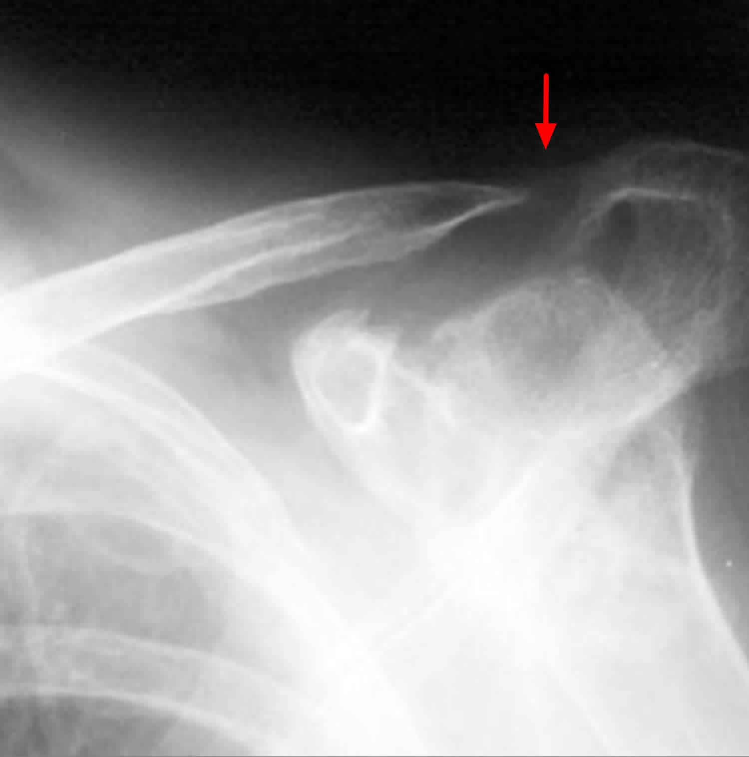
Diagnostic Procedures and Imaging
When you take a child to the emergency department for a suspected buckle fracture radius, the physician will perform a physical examination followed by an X-ray. It is important to note that because the fracture is a compression rather than a gap, it can sometimes be difficult to see on an X-ray unless the image is taken from exactly the right angle.
Radiologists look for a small "kink" or cortical disruption on the distal radius. Sometimes, a subtle thickening of the cortex is the only diagnostic clue. If the initial X-ray is inconclusive but the clinical suspicion remains high, doctors may recommend a follow-up visit or immobilization as a precautionary measure.
| Feature | Buckle Fracture (Torus) | Greenstick Fracture |
|---|---|---|
| Bone Integrity | Compressed/Buckled | Bent with partial break |
| Stability | Very Stable | Less Stable |
| Healing Time | Short (3–4 weeks) | Longer (6+ weeks) |
Treatment and Management Protocols
The treatment for a buckle fracture radius is generally straightforward and conservative. Because the injury is inherently stable, aggressive surgical intervention is almost never required. The primary goal is to provide pain relief and protect the bone while the natural repair process takes place.
- Immobilization: Most physicians will apply a removable splint or a short-arm cast to prevent further irritation.
- Pain Management: Over-the-counter pain relievers, such as ibuprofen or acetaminophen, are typically recommended, provided there are no contraindications.
- Follow-up Care: A follow-up visit is usually scheduled a few weeks later to assess healing and ensure there is no secondary displacement.
- Activity Restriction: Children are usually advised to avoid contact sports or high-impact activities for the duration of the healing period to prevent re-injury.
⚠️ Note: Always follow the specific advice of the treating physician regarding the duration of splint wear, as removing it too early can lead to persistent pain or improper healing.
Recovery and Return to Normal Activity
Recovery from a buckle fracture radius is typically very positive. Because the bone is not fully disrupted, the remodeling process is highly efficient in children. Within three to four weeks, most children can return to normal activities. During the recovery phase, it is common to notice that the wrist might feel slightly stiff after the splint is removed.
Physical therapy is rarely needed for this type of fracture. Gentle, active range-of-motion exercises—such as rotating the wrist or making a fist—are usually sufficient to regain full function. Parents should watch for any new pain or deformity after the splint is removed and consult their pediatrician if they have concerns.
Preventing Future Pediatric Injuries
While accidents are a natural part of childhood, certain environmental factors can be mitigated to reduce the risk of upper extremity trauma. Ensuring that playground equipment is well-maintained and that children wear appropriate protective gear when engaging in sports like skateboarding, cycling, or rollerblading can significantly decrease the likelihood of a buckle fracture radius.
Additionally, teaching children the "tuck and roll" technique or how to fall safely (trying to land on their feet rather than bracing with straight arms) can sometimes help prevent the specific type of impact that leads to forearm buckling. Encouraging bone health through adequate calcium and vitamin D intake is also a foundational aspect of pediatric orthopedic health, ensuring that bones are as resilient as possible against everyday impacts.
Ultimately, a buckle fracture radius is a manageable injury that typically resolves without long-term complications. By recognizing the signs early, seeking timely medical evaluation, and adhering to the prescribed period of immobilization, parents can ensure their child makes a full and swift recovery. While seeing a child in pain is distressing, these injuries serve as a reminder of the incredible healing capacity of the developing pediatric skeleton. Keeping the affected area protected and following clinical guidance remains the gold standard for restoring comfort and mobility to your child’s wrist.
Related Terms:
- buckle fracture radius radiology
- buckle fracture rch
- buckle fracture radius treatment
- buckle fracture radius children
- buckle fracture radius xray
- greenstick fracture radius