The human hand is a masterpiece of biological engineering, capable of performing tasks ranging from the most delicate surgical maneuvers to the exertion of significant grip strength. Understanding the anatomy of the hand wrist is essential for anyone interested in sports science, physical therapy, or simply maintaining long-term joint health. The wrist acts as the vital bridge between the forearm and the hand, providing a stable yet flexible foundation for the complex movements of our fingers and thumb. By exploring the interconnected systems of bones, ligaments, tendons, and nerves, we can better appreciate how this intricate region functions and why it is so susceptible to repetitive strain injuries.
The Skeletal Framework

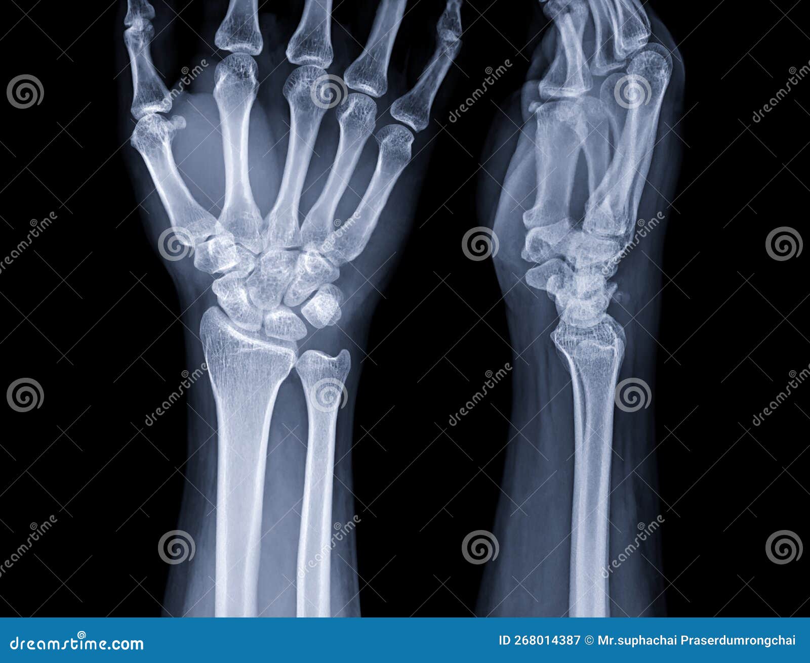
The anatomy of the hand wrist is built upon a complex arrangement of bones that allow for both stability and multi-axial movement. The wrist itself is comprised of the distal radius and ulna of the forearm, which articulate with the proximal row of carpal bones. There are eight carpal bones arranged in two rows, which act like a ball-bearing system to facilitate wrist flexion, extension, and rotation.
The structure of the hand bones is categorized as follows:
- Carpals: Eight small bones (scaphoid, lunate, triquetrum, pisiform, trapezium, trapezoid, capitate, hamate) located in the wrist.
- Metacarpals: Five long bones that form the palm of the hand.
- Phalanges: The 14 bones that make up the fingers and thumb; the thumb has two phalanges, while each finger has three.
Ligaments and Joint Stability
Because the carpal bones are small and highly mobile, they rely heavily on a dense network of ligaments to keep them in place. These tough, fibrous bands connect bone to bone, ensuring that the joints do not dislocate during movement. The scapholunate ligament is particularly critical; when this ligament is injured, it often leads to significant wrist instability and chronic pain.
The following table summarizes the primary functional components found within the wrist and hand:
| Component | Primary Function |
|---|---|
| Carpal Bones | Provide a flexible base for wrist movement |
| Metacarpals | Form the arch and palm for gripping |
| Collateral Ligaments | Provide side-to-side stability for fingers |
| Flexor Tendons | Allow fingers to curl and grasp |
| Extensor Tendons | Allow fingers to straighten or extend |
The Role of Tendons and Muscles
While the bones provide the structure, the tendons act as the cables that move the hand. In the anatomy of the hand wrist, these tendons pass through a tight channel known as the carpal tunnel, which is covered by the transverse carpal ligament. The extrinsic muscles, located in the forearm, send long tendons down into the hand, allowing for high-power movements. Conversely, the intrinsic muscles—those located entirely within the hand—allow for the fine, dexterous movements required for tasks like writing or threading a needle.
Common issues often arise when these tendons become inflamed, a condition frequently referred to as tenosynovitis. When the tendons are overworked, the protective sheaths surrounding them can swell, leading to discomfort and reduced range of motion.
⚠️ Note: Maintaining proper ergonomics at your workspace can significantly reduce the repetitive strain placed on these tendons, preventing chronic conditions like carpal tunnel syndrome.
Nerve Pathways and Sensory Feedback
The dexterity of the hand is entirely dependent on the complex network of nerves that run from the neck, down the arm, and into the fingers. The three primary nerves responsible for hand function are:
- Median Nerve: Passes through the carpal tunnel and provides sensation to the thumb, index, middle, and half of the ring finger.
- Ulnar Nerve: Responsible for the sensation in the little finger and the ring finger, as well as powering many of the intrinsic muscles of the hand.
- Radial Nerve: Primarily provides sensation to the back of the hand and assists in the extension of the wrist and fingers.
Movement Patterns and Biomechanics
Analyzing the anatomy of the hand wrist reveals how we achieve such a high degree of precision. The wrist joint allows for flexion (bending toward the palm), extension (bending toward the back of the hand), ulnar deviation (bending toward the little finger), and radial deviation (bending toward the thumb). This range of motion is further augmented by the carpometacarpal joint of the thumb, which is a saddle joint allowing for opposition—the ability to touch the thumb to each fingertip—a feature that distinguishes human manual capabilities from most other primates.
When studying the mechanics, it is important to observe how the bones, muscles, and nerves interact dynamically during activity. Even a slight misalignment in the carpal bones can create a domino effect that alters the entire kinetic chain of the upper limb.
💡 Note: Regular stretching and mobility exercises, specifically targeted at the forearm flexors and extensors, can help preserve the natural mobility of the wrist joint as you age.
Preventing Injury and Promoting Recovery
Given the complexity of the anatomy of the hand wrist, it is prone to various injuries, including fractures of the scaphoid bone, ligament tears, and nerve entrapments. Prevention strategies often center on strengthening the supporting muscles and ensuring that repetitive motions are punctuated by adequate rest periods. In cases of injury, recovery often involves immobilization or physical therapy designed to retrain the intrinsic muscles of the hand and restore the natural glide of the tendons through their sheaths.
Clinical assessments of the wrist often involve testing for specific signs of inflammation or nerve compression. By monitoring grip strength and range of motion, therapists can determine if the underlying structures are healing correctly. It is always best to consult with a medical professional if you experience persistent tingling, numbness, or weakness in the hand, as these are often indicators that the nerve pathways are under pressure.
The intricate design of the hand and wrist serves as a testament to the evolutionary necessity of touch and manipulation. By respecting the anatomical boundaries of these structures, we can ensure that our hands remain functional and strong throughout our lives. Whether through daily movement or specialized exercises, maintaining the health of the joints, nerves, and soft tissues in this region is fundamental to our overall physical interaction with the world. As we gain a deeper awareness of the biological systems at play, we are better equipped to protect these vital appendages from harm and support their recovery should injury occur.
Related Terms:
- muscles of the wrist diagram
- hand wrist anatomy diagram
- hand and wrist diagram
- left wrist muscle anatomy picture
- hand and wrist muscle anatomy
- fingers and wrists diagram